
Corneal topography has undergone remarkable technological evolution, yet its clinical value still depends heavily on how images are interpreted. In a recent overview published in ophta (3/2025), Torres-Netto, Hillen, and Hafezi revisit the fundamental principles of corneal topography and tomography, emphasizing that accurate diagnosis begins not with advanced indices, but with correct interpretation of basic maps
Modern imaging devices—ranging from Scheimpflug-based systems to anterior-segment OCT—provide increasingly detailed information about corneal shape and thickness. However, the authors stress that these modalities interact differently with corneal tissue. Blue-light systems are more sensitive to corneal transparency and scatter, whereas near-infrared OCT is less affected by optical haze. Understanding these differences is essential to avoid misinterpretation when comparing data across platforms.
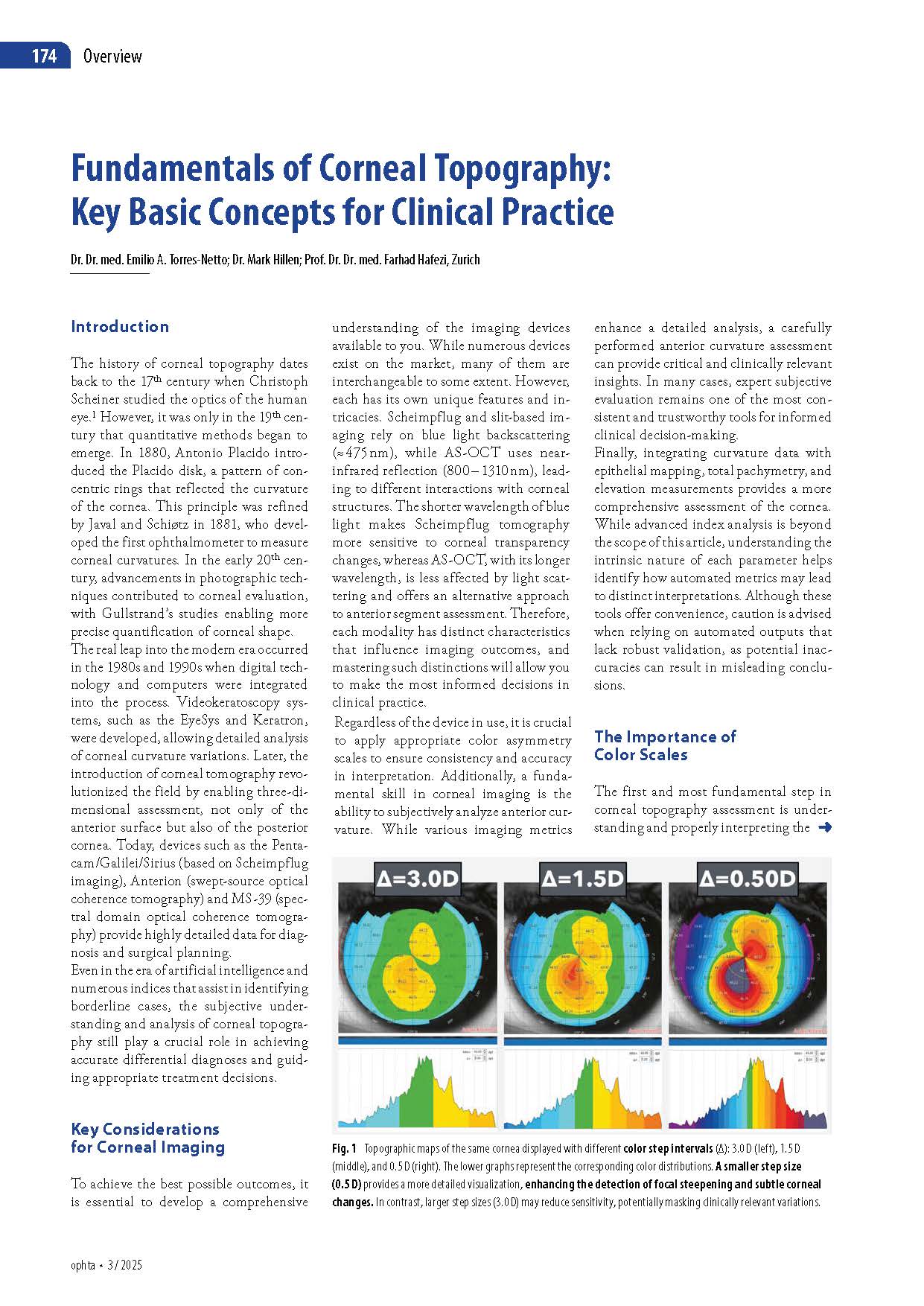
One of the most critical yet frequently overlooked factors is the use of color scales. The same cornea can appear markedly different depending on the chosen color step interval. Large steps may mask subtle focal steepening, while smaller steps enhance sensitivity to early ectatic changes. The authors recommend fixed color scales—rather than relative scales—to ensure reliable longitudinal comparison and reduce the risk of false normalization over time.
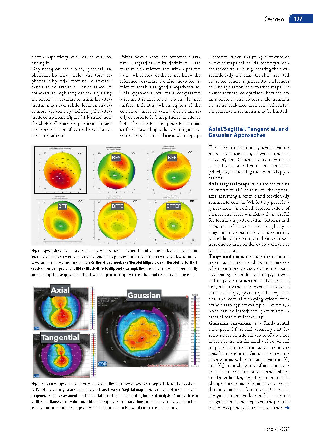
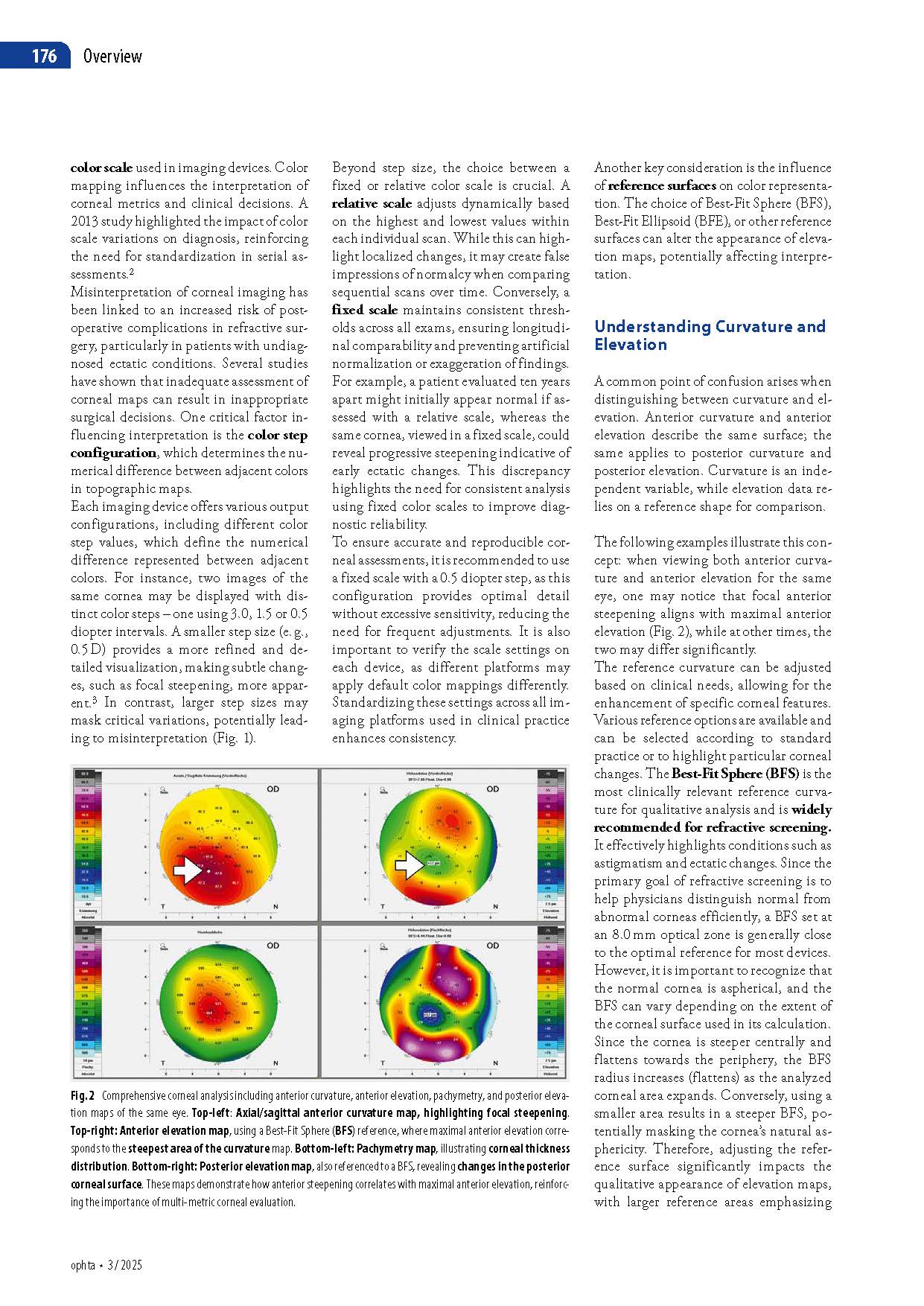
The article also clarifies the often-confused relationship between curvature and elevation. Although they describe the same corneal surface, elevation maps depend on the chosen reference shape, such as a best-fit sphere or ellipsoid. Changes in reference diameter or geometry can significantly alter map appearance, underlining the importance of consistency when monitoring patients over time.
Different curvature maps serve distinct clinical purposes. Axial (sagittal) maps provide a smoothed overview useful for refractive screening, while tangential maps are more sensitive to localized irregularities and early ectatic changes. Gaussian curvature, although less commonly used, offers additional insight into highly irregular corneas by describing intrinsic surface curvature independent of orientation.
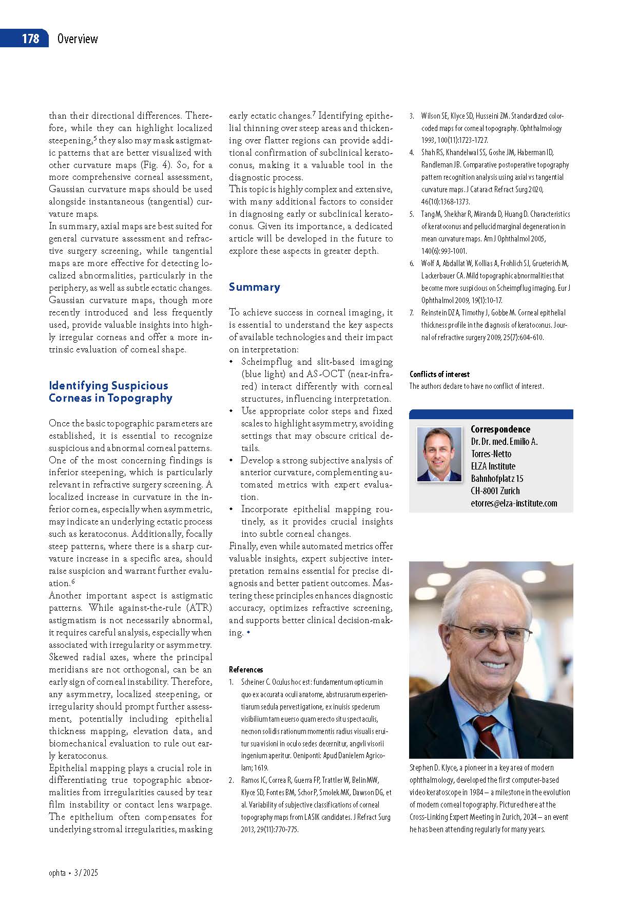
Finally, the authors highlight the importance of epithelial thickness mapping. Because the epithelium can partially mask underlying stromal irregularities, epithelial compensation patterns may reveal early keratoconus before conventional topography becomes clearly abnormal.
The overarching message is clear: while automated indices and artificial intelligence tools continue to evolve, expert subjective analysis remains indispensable. Mastery of color scales, reference surfaces, and basic curvature principles is essential for accurate diagnosis, safer refractive screening, and informed clinical decision-making in daily practice.