The human crystalline lens is an optical marvel that adjusts focus through accommodation, changing shape to shift focus. With age, this flexibility diminishes, leading to presbyopia. Understanding both how the lens deforms and how its optical properties shift is essential for advancing diagnostics and therapeutic design. A new human lens OCT study offers this insight by using optical coherence tomography to simultaneously map lens optics and mechanics.
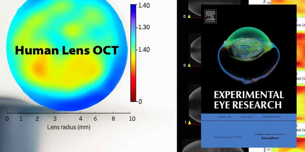
A recent study by Sabine Kling, Vahoura Tahsini, and Farhad Hafezi, published in Experimental Eye Research, introduces a breakthrough human lens OCT approach. Using high-resolution optical coherence tomography (OCT) and advanced phase-based signal processing, the team was able to map – simultaneously and in real time – both the gradient refractive index and the mechanical strain within the human lens under accommodative stress.
Human lens OCT study design
Six healthy adults (24–45 years) underwent OCT imaging under three accommodative demands: none, –2 diopters, and –4 diopters. Advanced signal-processing enabled pixel-level mapping of refractive index, while real-time micro‑fluctuation analysis yielded measures of:
-
Instantaneous strain (rate of deformation)
-
Accumulated strain (total deformation over time)
This dual mapping extends Dr Tahsini’s earlier OCE-based investigations into lens mechanics by adding simultaneous optical mapping to the biomechanical profiling portfolio.
Key findings
-
-
A pronounced axial gradient refractive index, with highest values in the posterior lens.
-
Refractive index proved stable across age and accommodative states in participants under 45.
-
Instantaneous strain decreased with age, indicating slower deformation in older lenses.
-
Accumulated strain rose with increasing accommodative demand.
-
No direct spatial correlation was observed between refractive index distribution and mechanical strain.
-
Why human lens OCT matters
This technique provides patient-specific maps of both optical and mechanical lens behavior, complementing this research group’s viscoelastic estimations of the lens and the infrastructure of SSI-based corneal stiffness mapping. Together, these methods deepen understanding of ocular tissue function with age and refractive state.
Potential applications include:
-
Early diagnostics for presbyopia
-
Personalized refractive treatment planning
-
Modeling emmetropization and myopization dynamics
By integrating OCT with biomechanical analysis, human lens OCT emerges as a translational tool for vision science, bridging optics and mechanics in a clinical context.
Reference
Kling S, Tahsini V, Hafezi F. Dynamic in vivo mapping of the gradient refractive index and strain distribution of the human lens under accommodative stress. Experimental Eye Research. 2025;255:110332. [PubMed]