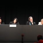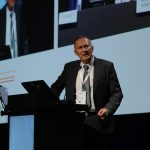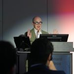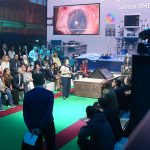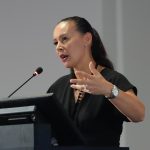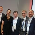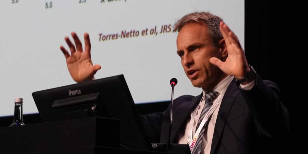
For decades, advanced keratoconus has ended in the same place. When nothing else works, the surgeon replaces the cornea with a full-thickness donor graft, a penetrating keratoplasty. A session at the 44th ESCRS Congress in London put a deadline on that assumption. Can full-thickness grafts for keratoconus disappear by 2040? Five surgeons answered with corneal transplant alternatives that stabilise, regularise or rebuild the cornea instead of replacing it. Three of the five work at the ELZA Institute.
Keratoconus thins the cornea, the clear window at the front of the eye, and lets it bulge into an irregular cone. Vision blurs and distorts, and glasses stop helping. Corneal cross-linking (CXL) stiffens the cornea with riboflavin drops and ultraviolet light, and it halts that progression in most patients. However, cross-linking was not designed to undo the shape the disease leaves behind. It has also been withheld from corneas that are already too thin.
Why corneal transplant alternatives matter in keratoconus
A full-thickness graft works, but the price is high. It carries a lifelong risk of rejection and a slow visual recovery. It also depends on donor tissue, which is scarce almost everywhere. Every technique in the session therefore shared one ambition. Take less from the donor pool, and give the patient back a cornea that is both stable and better shaped.
Bowman layer grafts, stability without sharper vision
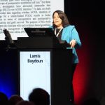
Dr. Lamis Baydoun opened with the caveat most speakers keep for the end. Her technique does not make anyone see better. “You can only tell your patient their contact lens vision will be perfect,” she said. “Nothing will be different after that.”
The procedure was first published in 2014. It implants an isolated Bowman layer graft, a single acellular layer taken from a donor cornea, into a pocket inside the patient’s own stroma. The graft flattens and stabilises corneas that are still progressing but are too thin for cross-linking. The tissue economy is striking. One donor cornea can yield a Descemet membrane endothelial keratoplasty (DMEK) graft, a deep anterior lamellar keratoplasty (DALK) button and a Bowman layer graft. Three patients are treated from tissue that once served one.
In her larger cohort, roughly 90 per cent of eyes held their ground against further progression. Higher-order and spherical aberration fell measurably, although visual acuity stayed flat. The obstacle is the surgery itself. Harvesting the graft is a knife-edge manoeuvre, because too thin and it tears, too thick and stroma is left behind. That difficulty pushed her team towards a simpler variant, the Bowman layer onlay, performed entirely outside the eye. Five early eyes flattened cleanly. A larger series, however, showed some dislocation and incomplete healing. As she put it, “We really have to do some more research” before this replaces anything.
ELZA-PACE, reshaping the cornea without donor tissue
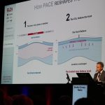
Prof. Farhad Hafezi presented the only genuinely donor-free option in the session. He also reported its most striking number, up to 20 D of regularisation in selected cases. No donor tissue is added, and no stroma is removed. The protocol is ELZA-PACE, a customised epi-on cross-linking treatment assisted by phototherapeutic keratectomy.
Its mechanism rests on one small, deliberately placed epithelium-off window of 2.5 mm by 3.5 mm. That window concentrates four effects exactly where the cone needs them. More oxygen reaches the stroma, riboflavin penetrates better, UV exposure rises, and the epithelium resists less. The result, Prof. Hafezi said, is coupling. The cone flattens, the surrounding tissue steepens, and the surface regularises using the patient’s own cornea.
The underlying epi-on protocol already has laboratory support. A 2025 study found that it matched accelerated epithelium-off cross-linking on both demarcation depth and biomechanical strengthening. The clinical study confirming the effect in patients is in press.
The preliminary one-year clinical results are cautious but encouraging. Forty per cent of the cohort dropped at least one disease stage. No eye progressed further, and no eye lost lines of vision. There was no clinically significant haze either, despite a fluence nearly triple that of a standard protocol. Because nothing is removed except a small window of epithelium, Prof. Hafezi treats keratoconus in patients as young as 13. At that age he would use neither an excimer laser nor a donor graft. The current ceiling is regulatory rather than clinical, since the riboflavin formulation has been awaiting approval for more than two years.
Ring segments carved from donor cornea
Dr. Shady Awwad traced the path from the original corneal allogenic intrastromal ring segments (CAIRS), described in 2018, to a fully software-planned version cut with a femtosecond laser and first published in 2023. These segments are carved from donor corneal tissue rather than manufactured from plastic. The innovation therefore lies in swapping the material, not in removing the donor from the equation.
CAIRS shares the host cornea’s refractive index, and nutrients diffuse through it. As a result, it tolerates placement that rigid polymethyl methacrylate (PMMA) segments cannot. Optical zones can be as small as 4 mm, at shallower depths, with little of the glare that pushes plastic segments to 5 mm or wider.
His rule of thumb concerns asymmetry. If the astigmatic and comatic axes differ by more than 60 degrees, a symmetric segment risks worsening one problem while fixing the other. Asymmetric segments separate the two. In his series, that approach flattened K-max, a measure of how steep the cornea is, by roughly 5 D. It also produced meaningful reductions in coma, a distortion that smears light into a comet-like tail. A propensity-matched retrospective analysis, which he described as the only comparison of its kind so far, showed slightly better corrected visual acuity than PMMA segments at equivalent flattening.
The complications were real but manageable. They included three cases of tunnel-roof melt, epithelial erosion in contact lens wearers that resolved when the lens was removed, and one or two rejections. His closing warning was pointed. Without a shared registry, CAIRS risks repeating twenty years of failure to produce an evidence-based nomogram for plastic segments.
ECO-CAIRS, cross-linking the graft before it goes in
Dr. Emilio Torres-Netto took cross-linking outside the eye altogether. In ECO-CAIRS, the donor ring segments are cross-linked at an ultra-high fluence of 30 J/cm² before implantation. The result in theatre is a graft that is firmer and drier. It threads through the corneal tunnel more easily, and it swells less. No living cells remain in the segment, so the cross-linking step may also lower the risk of rejection. Dr. Torres-Netto was careful to note that this has not been established.
He also reported something the field cannot yet explain. CAIRS segments do not reproduce the pattern of biomechanical relaxation seen around PMMA rings, which suggests two genuinely different mechanisms. One case made the argument for donor tissue in a single image. A scar from a previously explanted PMMA ring sat exactly where a new segment had to go. That terrain would have been too fragile for a rigid ring, yet the flexible CAIRS graft threaded around it. Vision improved from 0.4 to 0.8 decimal visual acuity, roughly 20/50 to 20/25 on a Snellen chart.
SLAK, filling the cone instead of flattening around it
Prof. Emilio Pedrotti closed with the most structurally different idea. Instead of placing a ring around the cone, he puts tissue back inside it. Building on a technique first proposed by Prof. Leonardo Mastropasqua, he uses a positive meniscus lenticule, thicker in the centre. It is cut from a donor cornea and inserted into a deep stromal pocket to rebuild the thinned area.
The effect lands where the disease starts, because the posterior surface of the cornea regularises most. The anterior surface is more stubborn. Prof. Pedrotti therefore follows stromal lenticule additional keratoplasty (SLAK) with customised phototherapeutic keratectomy (PTK), a laser polishing step that removes the remaining irregularities. Together, he reported, the two produced a three-line gain in best-corrected spectacle vision. Corneal stiffness rises substantially after SLAK, dips slightly after PTK, and still finishes well above baseline. Crucially, nothing is foreclosed. A full keratoplasty remains available if SLAK underdelivers.
What still stands in the way
Taken together, the corneal transplant alternatives presented in London amounted to one donor-free technique and four that stretch a single donor cornea much further than a full transplant ever could. Some treat several patients from one donor eye. Others use smaller pieces, or place tissue in configurations that a rigid plastic segment cannot match. The honest verdict, though, was that donor corneas are not going anywhere. They are simply being asked to do more.
Three obstacles came up repeatedly. First, progression still has to be stopped before any reshaping technique is worth attempting. Second, the nomograms that tell a surgeon exactly what to implant, and where, remain largely unpublished. Third, long-term data are thin for every technique except the oldest. On the evidence presented in London, 2040 looks less like a deadline for the full-thickness graft. It looks more like a target the field is still working towards.
The ELZA Institute offers keratoconus diagnosis and treatment, including customised cross-linking and corneal surgery. Its surgeons also contribute to the research behind several of the techniques above. The 44th ESCRS Congress ran from 11–15 September 2026 at ExCeL London.
References
- van Dijk K, Parker J, Tong CM, et al. Midstromal isolated Bowman layer graft for reduction of advanced keratoconus. JAMA Ophthalmol. 2014;132(4):495–501. doi:10.1001/jamaophthalmol.2013.5841
- Lu NJ, Torres-Netto EA, Aydemir ME, et al. A transepithelial corneal cross-linking (CXL) protocol providing the same biomechanical strengthening as accelerated epithelium-off CXL. J Refract Surg. 2025;41(7):e724–e730. doi:10.3928/1081597X-20250515-09
- Jacob S, Patel SR, Agarwal A, et al. Corneal allogenic intrastromal ring segments (CAIRS) combined with corneal cross-linking for keratoconus. J Refract Surg. 2018;34(5):296–303. doi:10.3928/1081597X-20180223-01
- Bteich Y, Assaf JF, Gendy JE, et al. Asymmetric all-femtosecond laser-cut corneal allogenic intrastromal ring segments. J Refract Surg. 2023;39(12):856–862. doi:10.3928/1081597X-20231018-04
- Asfar KE, Bteich Y, Abou Mrad A, et al. Corneal allogenic intrastromal ring segments (CAIRS) versus synthetic segments, a single segment comparative analysis using propensity score matching. J Refract Surg. 2024;40(11):e863–e876. doi:10.3928/1081597X-20241002-02
- Hafezi F, Aydemir ME, Hillen M, et al. Extracorporeal optimization of corneal allogenic intrastromal ring segments (ECO-CAIRS) using ultra-high-fluence corneal cross-linking. J Refract Surg. 2025;41(11):e1233–e1239. doi:10.3928/1081597X-20250917-02
- Kollros L, Aydemir ME, Boote C, et al. Dehydration and rehydration behavior of ultra-high-fluence extracorporeal cross-linked corneal allogenic intrastromal ring segments (ECO-CAIRS). J Cataract Refract Surg. 2026, ePub ahead of print. doi:10.1097/j.jcrs.0000000000002015
- Mastropasqua L, Nubile M, Salgari N, Mastropasqua R. Femtosecond laser-assisted stromal lenticule addition keratoplasty for the treatment of advanced keratoconus. J Refract Surg. 2018;34(1):36–44. doi:10.3928/1081597X-20171004-04
- Perez M. Can full-thickness corneal transplants for keratoconus disappear by 2040. CAKE Magazine, 13 September 2026. CAKE Magazine session report