At the ELZA Institute in Zurich, we use advanced diagnostic tools and a team-based approach to detect keratoconus early – before your vision is affected. Early diagnosis gives you more options for treatment and helps protect your sight.
Keratoconus is a condition where the cornea becomes thinner and bulges outward, distorting your vision. It often develops slowly and may go unnoticed in its early stages. That’s why we rely on highly precise imaging and biomechanical testing to detect even the smallest changes in your cornea – long before symptoms appear. Keratoconus can be treated with corneal cross-linking (CXL), but CXL typically only halts disease progression – on its own, CXL cannot recover the vision that was lost before it was performed. This is why early detection and treatment is so important with keratoconus – to preserve as much vision as possible.
Our Diagnostic Tools: What We Use and Why
We don’t rely on just one test. Instead, we combine several advanced technologies to build a complete picture of your eye health.
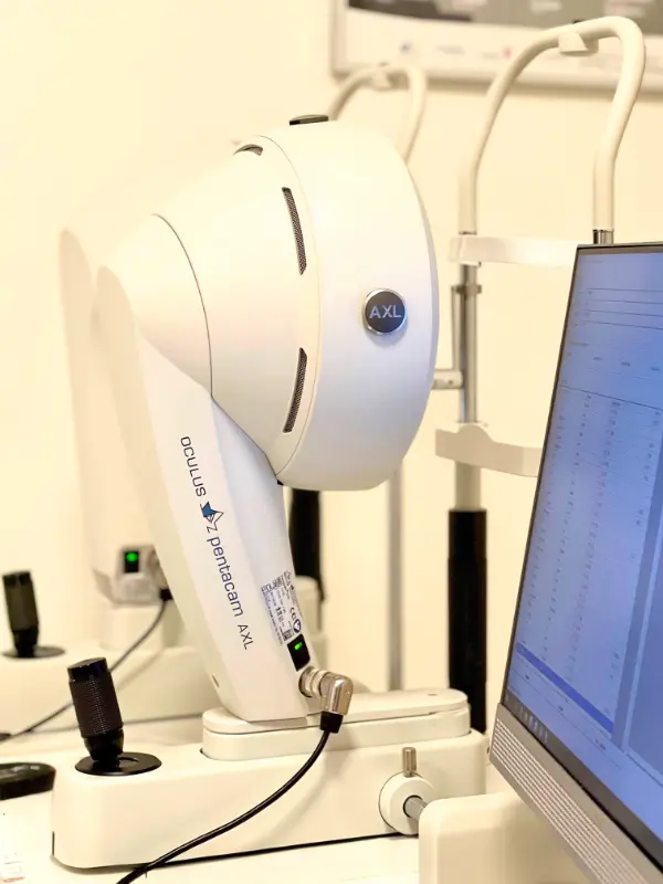
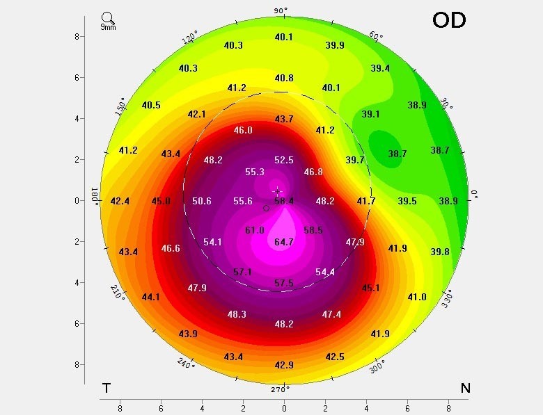
Pentacam
Pentacam: 3D Mapping for Early Detection
The Pentacam uses a rotating camera to capture a 3D image of your cornea, measuring its shape and thickness at thousands of points. This lets us detect keratoconus at a very early stage – even when vision still seems normal.
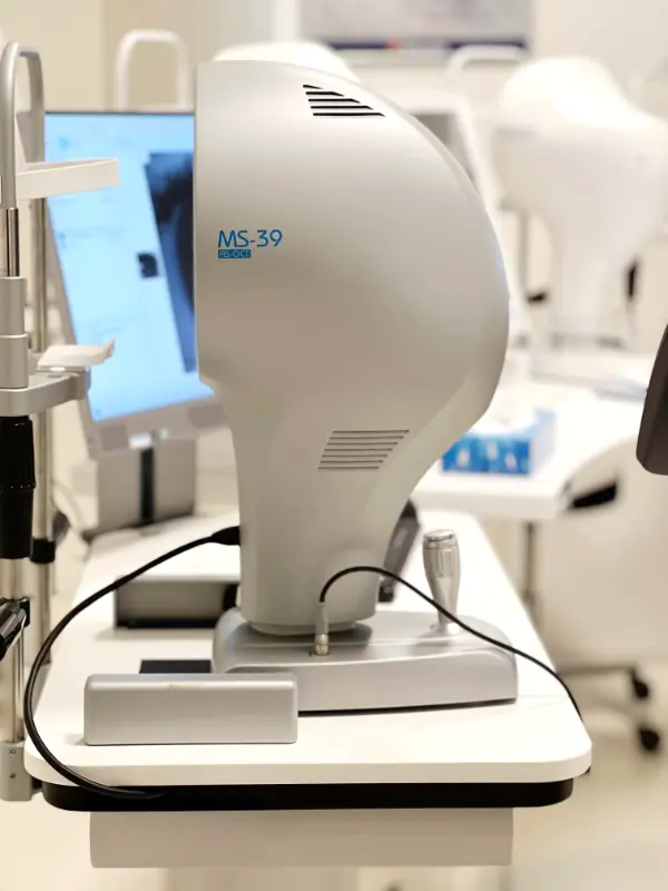
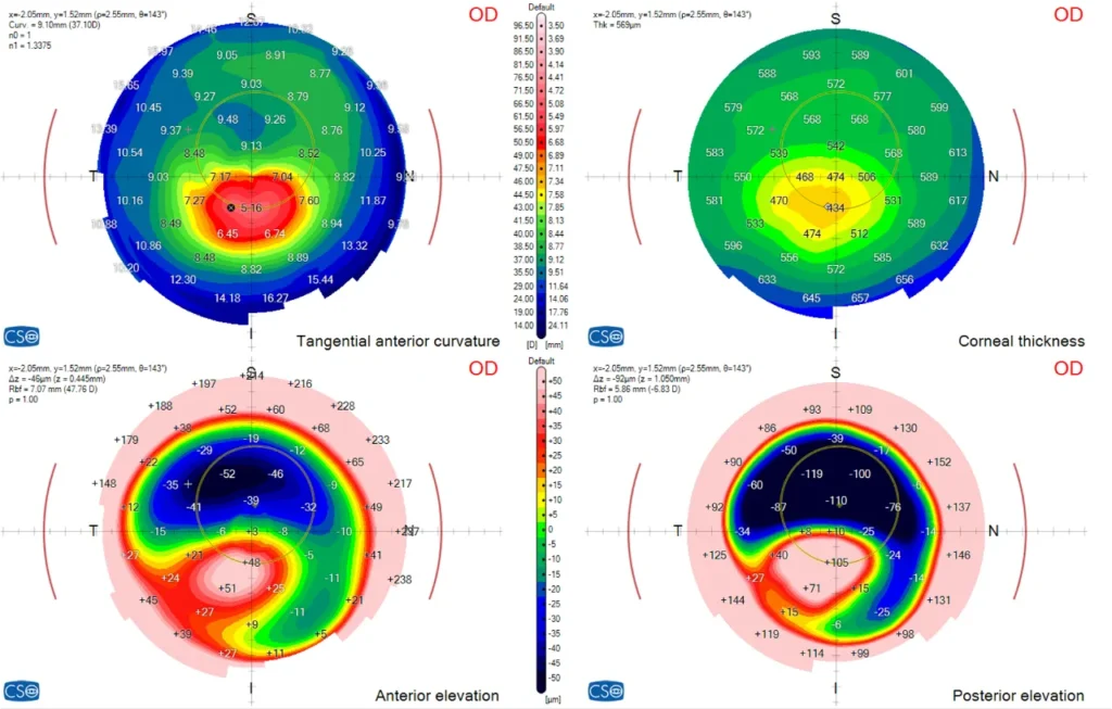
M–39
MS-39: High-Resolution Imaging and Corneal Structure
The MS-39 combines corneal topography with high-resolution OCT (optical coherence tomography). It gives us an extremely detailed image of the corneal layers, allowing us to detect subtle structural changes and monitor any progression over time.
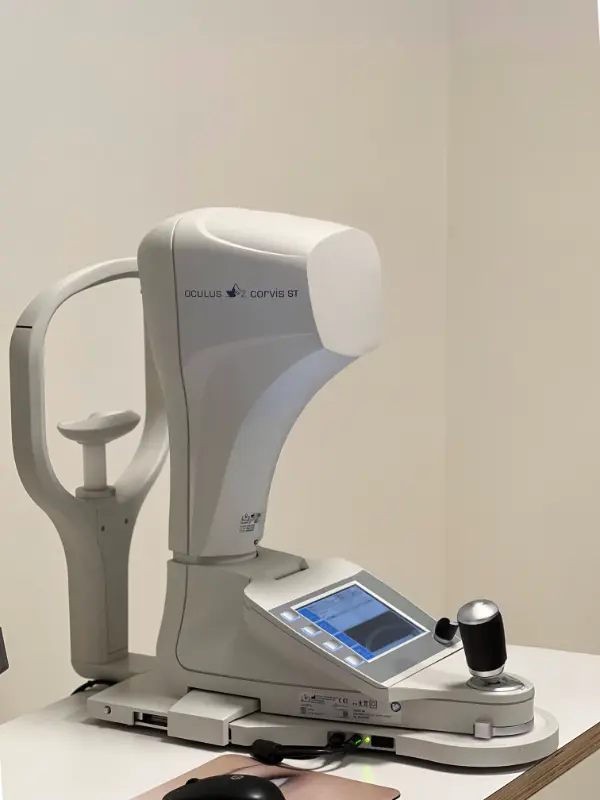
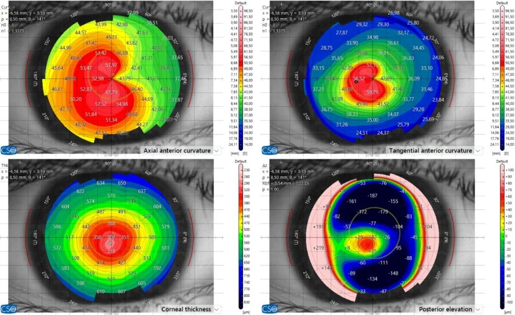
Corvis-ST
Corvis ST: Measuring Corneal Strength
Keratoconus isn’t just about shape, it’s also about strength. The Corvis ST measures how your cornea responds to a gentle puff of air, helping us assess its biomechanical stability. This information is critical for early diagnosis and determining whether treatments like cross-linking are needed.
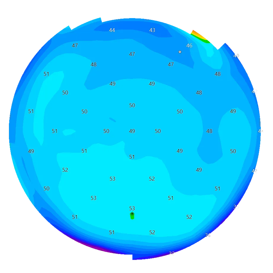
Normal Cornea
A normal healthy cornea is smooth and has the same thickness throughout. The shape of the cornea resembles a ball, with with the same amount of curvature in all directions.
Regular astigmatism
The cornea is more curved in one direction than the other – in other words, the cornea is shaped more like an egg than a ball. This shape distorts vision, but can also easily be corrected with glasses because the distortion is symmetric.
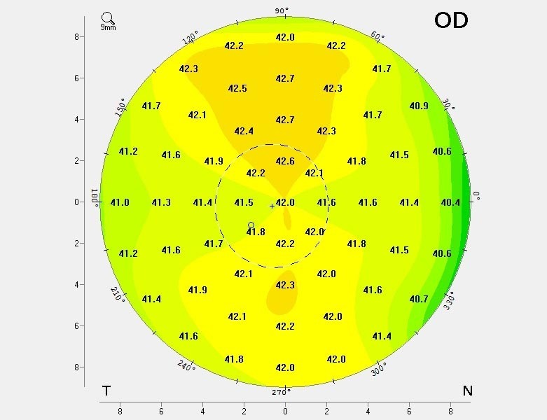
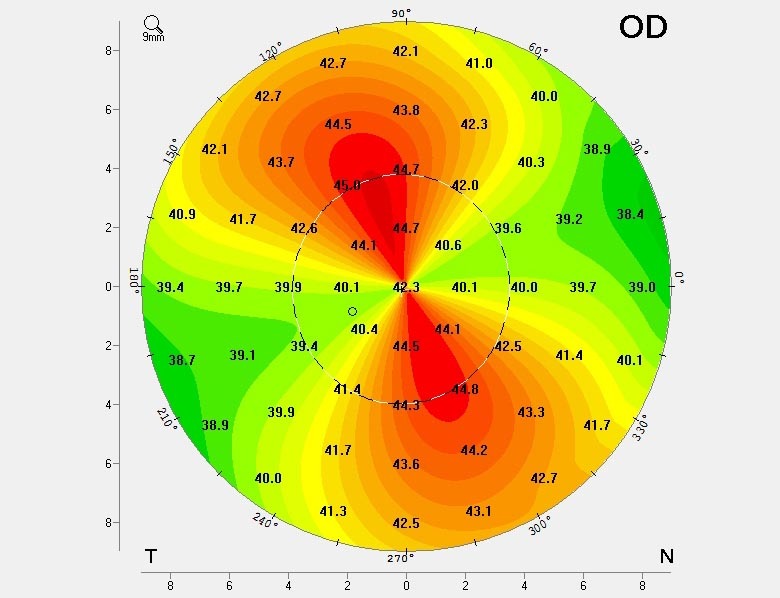
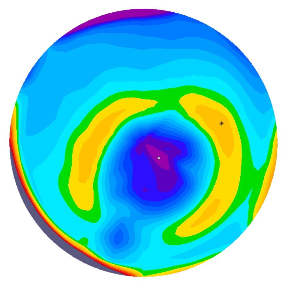
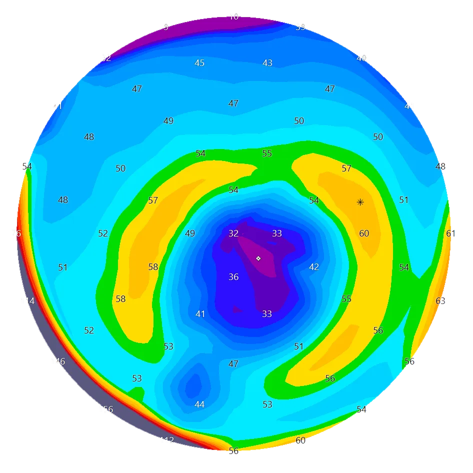
Keratoconus
This cornea is diseased and needs further examination and treatment. In the case shown in the image below, the cornea is flat in the upper part and markedly bulges out in the lower part (which is what’s known as the “cone”). Vision is distorted (people see double, triple and even quadruple images, and in many cases, glasses cannot restore decent visual acuity. Further, keratoconus is progressive and can lead to severe visual impairment, especially in children and adolescents.
Regular Monitoring Is Key
Keratoconus can change over time, especially in younger patients. At ELZA, we emphasize regular monitoring using the same advanced instruments. These follow-up tests allow us to track changes and step in with treatment before the condition worsens.
Tailored to You: Diagnosis and Treatment Planning
Because every eye is different, we combine your imaging results, visual tests, and clinical examination to develop a treatment plan specifically for you. This might include custom contact lenses, corneal cross-linking to stop progression, or other advanced procedures—all under one roof at our center in Zurich.
Trusted by Patients Worldwide
Our diagnostic technologies are matched by the quality of our team. ELZA is home to internationally recognized corneal specialists, led by Prof. Farhad Hafezi MD, PhD, FARVO, Dr. Emilio Torres-Netto, MD, PhD, FWCRS, and PD Dr. Lamis Baydoun, MD, PhD. Together, we’ve helped define global standards in keratoconus diagnosis and treatment—and we bring that same expertise to your individual care.
Pediatric Keratoconus: Special Attention for Young Eyes
Keratoconus tends to progress more rapidly in children and adolescents. That’s why we offer dedicated screening and treatment options for younger patients, giving them the best chance to preserve vision over the long term.
What You Can Expect
When you come to ELZA for a keratoconus assessment, you’ll receive:
A full diagnostic work-up using Pentacam, MS-39, and Corvis ST
Expert interpretation by cornea specialists
Personalized treatment recommendations
Follow-up monitoring using the same instruments to ensure consistency and precision
Your Vision. Our Priority.
Keratoconus can be challenging, but with early diagnosis and the right care, it doesn’t have to limit your life. At ELZA, we’re here to guide you every step of the way.
Topography and Tomography
Thanks to the latest technologies, we are now able to measure both the anterior and posterior surfaces of the cornea with extremely high precision. This enhanced level of accuracy allows us to detect abnormalities at very early stages, leading to faster and more reliable diagnoses. Conditions such as keratoconus or pellucid marginal degeneration (PMD) can now be identified much earlier than before, improving patient outcomes and enabling timely treatment.
In addition, this technology provides an extra level of safety when it comes to laser eye surgery. By offering a much more precise assessment of the cornea, it helps us determine with greater confidence whether a patient is a suitable candidate for procedures such as LASIK or PRK, significantly reducing the risk of complications.
Corneal biomechanics
Corneal biomechanics is another essential parameter in both the diagnosis and management of keratoconus, as well as in determining eligibility for refractive surgery. By assessing the biomechanical properties of the cornea, we gain valuable insight into how likely it is to deform or develop ectasia, a progressive corneal disease often associated with structural weakness.
A lower biomechanical resistance may indicate a higher risk of corneal instability, which is critical information when evaluating candidates for procedures like LASIK or PRK. This makes biomechanical assessment a key part of our preoperative screening process and long-term patient monitoring. Our group is actively involved in ongoing research in this field, contributing to advancements in understanding and improving diagnostic and predictive tools related to corneal biomechanics.
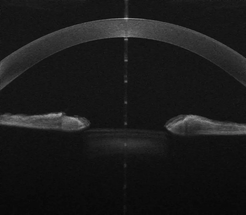
High-resoluion OCT Image
Modern anterior segment OCT technology offers ultra high-resolution imaging of the corneal tissue, allowing for incredibly detailed visualization of its structure. This level of precision represents a major advantage in the follow-up of corneal diseases, as well as post-operative monitoring. It enables clinicians to track subtle changes in the cornea over time with great accuracy.
One of the key benefits of this imaging is the ability to assess corneal scars, not only in terms of their presence, but also their depth and extent within the corneal layers. Such detailed information is crucial for evaluating the impact on visual quality and for planning future treatment strategies when needed.