For most of the history of corneal surgery, reshaping a cornea has meant taking part of it away. The closing session of the 44th ESCRS Congress in London started from the opposite premise. ECO-CAIRS keratoconus data were ELZA’s last contribution to the week, on Tuesday 15 September.
Reshaping a cornea by adding tissue rather than removing it
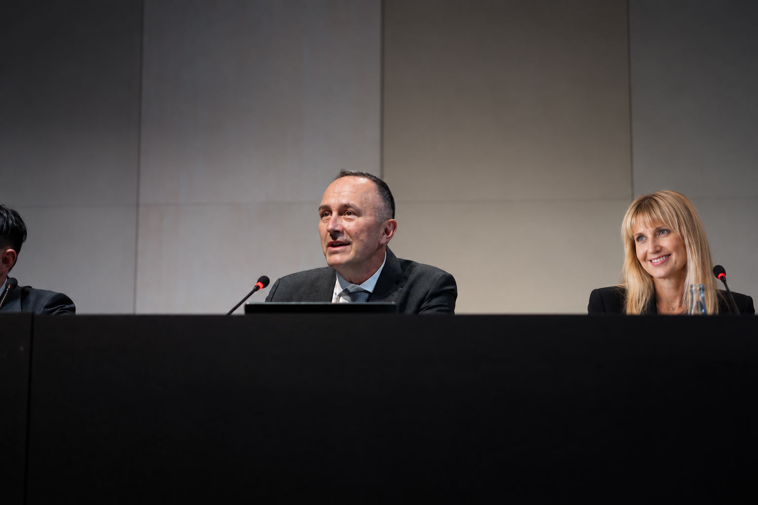
Prof. Farhad Hafezi co-chaired the free-paper session on corneal reshaping in keratoconus at 11:00, with Prof. Aylin Kilic. He presented in it at 11:35. The session’s theme reflects a real shift in the field. Laser refractive surgery works by ablation, removing stromal tissue to flatten the cornea. In an eye already weakened by keratoconus, however, removing tissue is the last thing anyone wants to do.
Corneal allogenic intrastromal ring segments, or CAIRS, take the other route. Arcs of donor corneal tissue are implanted into a channel in the patient’s own stroma. There they flatten the cone from within. Nothing is removed. The segments are human corneal tissue rather than plastic, so they also avoid some complications associated with synthetic rings.
What ECO-CAIRS keratoconus treatment adds to the technique
The ELZA modification cross-links the segments before they go in. Corneal cross-linking (CXL) uses riboflavin – vitamin B2 drops – and ultraviolet light to stiffen corneal collagen. Here it is applied to the donor segment outside the eye, at ultra-high fluence, rather than to the patient’s cornea. ELZA calls the result ECO-CAIRS.
Twenty eyes of twenty patients with stable keratoconus were included. Maximum keratometry ranged from 58.00 to 69.80 D. In addition, corneal thickness was at least 400 µm at 5 mm from the pupil centre. Segments were cut from donor corneas with a femtosecond laser. They were then treated with either 30 or 60 J/cm², and implanted without air-drying or prolonged dehydration.
Insertion was uneventful in every case. The cross-linked segments stayed stiff throughout implantation, instead of softening as surgeons handled them. Segment thickness fell by roughly half immediately after treatment. Partial re-expansion followed over the next few days, once the segment was inside the stroma.
Why a thinner, stiffer segment is useful
Three practical consequences follow. First, a stiffer segment gives the surgeon more control during insertion. Second, ultraviolet treatment at this dose leaves the segment acellular. What is implanted is therefore a stable collagen scaffold, rather than living donor tissue. Third, the segment temporarily loses about half its thickness. A larger-volume segment can therefore be threaded through the same channel, which may increase the topographic effect once it re-expands.
The authors are explicit that this is an early series. Twenty eyes describe surgical behaviour, not refractive outcomes over time. Further studies are needed to separate the contribution of segment volume from that of the stiffness itself. What the ECO-CAIRS keratoconus data support is handling and structural stability, which is what the abstract claims.
How the week fitted together
Read across the five days, ELZA’s London programme kept returning to one idea. Friday and Saturday made the case for measuring how a cornea behaves, rather than only how it looks. Sunday supplied the long-term evidence that cross-linking holds at ten years. It also supplied the honest counterpart, in a series of cases where the treatment went wrong. Monday showed how much of keratoconus is still managed optically, with lenses rather than surgery. Tuesday closed on tissue that is added rather than subtracted.
The ELZA Institute performs ECO-CAIRS and corneal cross-linking for keratoconus at its clinics in Dietikon and Zurich. Session details are published by the European Society of Cataract and Refractive Surgeons.