Not every keratoconic eye needs surgery. Many are managed with a lens that vaults clear over the irregular cornea. Scleral lenses keratoconus care of that kind was ELZA’s most quantified contribution to Monday 14 September at the 44th ESCRS Congress in London.
What scleral lenses keratoconus patients wear actually do
Keratoconus distorts the front surface of the eye, and a distorted surface bends light unevenly. The result is not simply blur that stronger glasses can fix. It is higher-order aberration, an optical error with no spectacle equivalent. A mini-scleral lens rests on the white of the eye and arches over the cornea. A reservoir of fluid sits underneath. Optically, the eye’s irregular front surface is replaced by the smooth front surface of the lens.
Léonard Kollros presented the measurements at 14:35, in the free-paper session on management of advanced keratoconus and complex cornea. Twenty-two eyes of twelve adults with stable keratoconus were assessed with and without their habitual lenses. Each had spent at least ten days out of lens wear beforehand. In addition, seventeen of the eyes had previously had cross-linking.
Total higher-order aberrations fell from a median of 1.08 µm without the lens to 0.51 µm with it. Vertical coma dominates in keratoconus, and it dropped from −1.23 µm to −0.03 µm. That is a mean correction of 97.3%. High-contrast visual acuity improved from 0.21 logMAR to −0.06 logMAR. Scores on the NEI VFQ-25 quality-of-life questionnaire rose from a median of 62.4 to 83.8.
That last number is the one worth pausing on. Optical measurements are easy to collect and easy to over-interpret. A validated questionnaire, by contrast, asks patients whether daily life improved. Here both moved together. The study is small and single-centre, however, and the eyes were stable rather than progressing. As evidence for scleral lenses keratoconus management, that is useful but modest.
Turning a research idea into something that exists
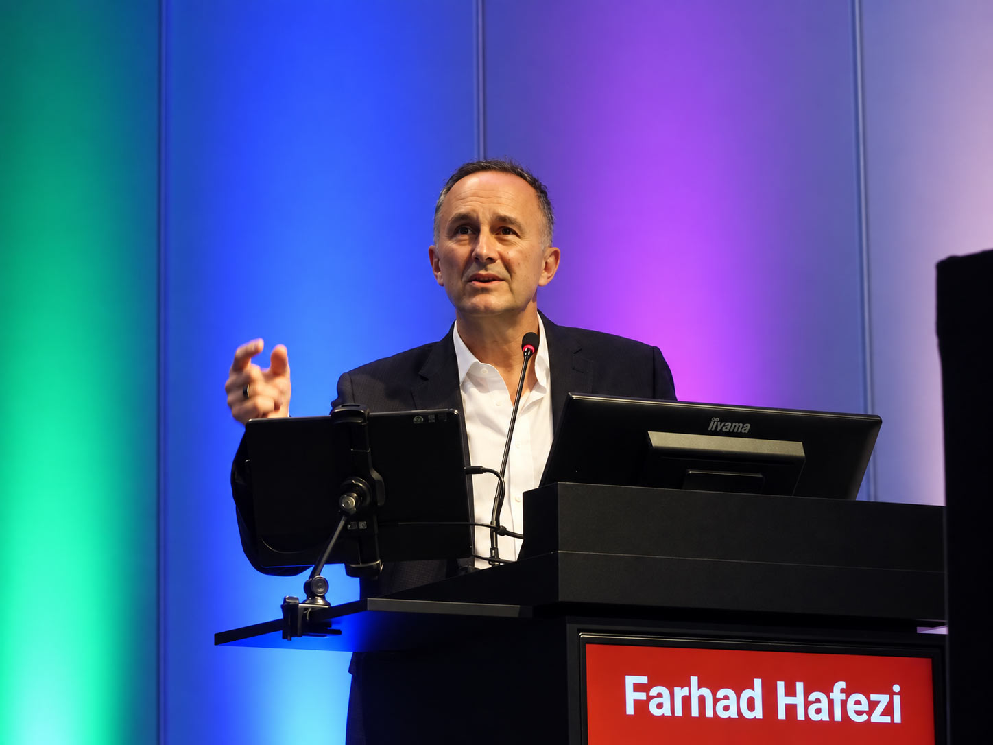
At 14:30, in Capital Suite Room 2-4, the ESCRS Research Workshop asked a question most methods courses skip. How do you tell an idea worth pursuing from one that is not? The workshop, I have a great idea… now what?, was chaired by Prof. Farhad Hafezi. Nikki Hafezi opened it. She works on public health, policy and strategic management in ophthalmology.
Her session covered identifying an unmet need, and then validating it before committing years to it. The workshop then followed a clinical idea the whole way to a product. It covered patents and ownership, prototype to certified device, and who funds each stage. It also set out why drug development plays by different rules. Open discussion closed the session.
Imaging the cornea, and measuring how it moves
At 17:30, Dr Emilio A. Torres-Netto taught optical coherence tomography elastography. His slot sat within an instructional course on multimodal anterior segment imaging, in Capital Suite Room 9/10. Conventional imaging maps the shape of the cornea. Elastography attempts to map its stiffness directly. It therefore belongs to the same shift that ran through ELZA’s Saturday programme: measuring how tissue behaves, not only how it looks.
Glaucoma surgery in combination
Five minutes later, in Capital Suite Room 14-16, Prof. Kaweh Mansouri spoke in a course on combined deep sclerectomy and cataract surgery. He compared it with other combined glaucoma procedures. Deep sclerectomy creates a drainage route for fluid inside the eye without entering the anterior chamber. Because of that, it carries a different risk profile from trabeculectomy.
Corneal transplantation in the Arena
Earlier in the morning, at 09:55, Dr Lamis Baydoun demonstrated the unrolling technique in the Arena’s DMEK session. A DMEK graft is a donor membrane a few cells thick, and because it is so thin it curls on itself in fluid. Unrolling it is the step that most often separates a smooth case from a difficult one. It is also taught almost entirely by demonstration.
The ELZA Institute manages keratoconus with optical as well as surgical approaches. It performs corneal surgery and glaucoma surgery at its clinics in Dietikon and Zurich. Session details are published by the European Society of Cataract and Refractive Surgeons.