The Middle East Africa Council of Ophthalmology (MEACO) Congress is one of the highlights of our congress calendar each year. It attracts some of the biggest names in ophthalmology, and the standard of the presentations and the quality of debate is as good as any conference in our specialty.
I was honoured to be invited to give a series of talks during this year’s MEACO congress, held in the Hilton Dead Sea & Spa Hotel in Amman, Jordan.
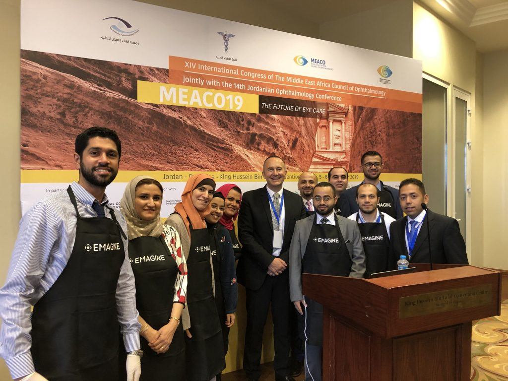
[foogallery id=”16605″]
Individualized CXL for thin corneas
My first task was to participate in the MEACO “Experts’ Top Tips” session, where I gave a presentation on CXL and Thin Corneas, reviewed the history of performing CXL in thin corneas, followed by the work performed by our research group at the University of Zurich that examined the essential components of the cross-linking photochemical reaction: chromophore availability, UV light intensity, exposure time and the biological response, how we combined each of these factors to a model, and how we’re using that model, combined with measurements of corneal thickness, to tailor cross-linking protocols to each person’s individual corneal thickness – and how this can be used to treat thin corneas, without resorting to swelling the cornea with hypo-osmolar riboflavin or other approaches like contact lens-assisted cross-linking.
PACK-CXL
My next talk was in the infectious keratitis session, where I spoke about using CXL to treat infectious keratitis (PACK-CXL). During cross-linking, an overwhelming amount of Reactive Oxygen Species (ROS) are created when UV light photo-activates the riboflavin that has been applied to the cornea. This not only cross-links the collagen molecules in the stroma, strengthening the cornea and increasing its resistance to ectasia, but also catabolic enzymes, such as those produced by inflammatory processes – or invading pathogens. But the ROS serve a more important purpose in PACK-CXL: they damage the cell walls of invading pathogens and intercalate with their DNA. CXL – whether it’s used to treat ectasia or infection renders the cornea sterile. And so I was happy to review what we currently know about PACK-CXL, from the basic science, to the latest updates from large-scale clinical evaluations of PACK-CXL for the treatment of keratitis. CXL is now possible with portable, rechargeable, slit lamp-mounted CXL devices – and this represents a phase shift from current clinical practice. CXL is currently performed in sterile, expensive operating rooms. It doesn’t have to be. It can be performed anywhere there’s a slit lamp. And given slit lamps are ubiquitous – wherever there’s an eye doctor, there’s a slit lamp, this dramatically opens up the possibility of treating infectious keratitis with CXL to most parts of the world – not large hospitals in major population centres. PACK-CXL has been used to successfully resolve infectious keratitis without even the use of antimicrobial drugs – with a single treatment. This could prove to be increasingly valuable as antimicrobial drug resistance rises – and in parts of the world where access to these drugs is limited.
On Friday, I was involved in the MEACO “Controversies in CXL” session, where I spoke about “Epi-off CXL”. For me, there is no doubt in my mind that for now epi-off remains the gold standard of cross-linking: for CXL to work, riboflavin has to be in the corneal stroma, and the epithelium has to be removed for the riboflavin to get there. Attempts to force the riboflavin through by chemical or electrochemical means still result in poorer riboflavin penetration into the stroma, less effective cross-linking, and an increased probability of keratoconus progressing again later, and I remain unconvinced otherwise by published data to date – so in this talk, I reviewed the evidence base behind my conclusions. However, this might change in the near future, because more sophisticated epi-on protocols may help increase the efficacy of epi-on.
CXL at the Slit Lamp
I later spoke about cross-linking at the slit lamp – again, reviewing the reasons why CXL does not need a sterile OR to be performed as it sterilises the cornea, and the cost and availability advantages this approach brings – in particular, bringing effective, potentially “one-shot”, antimicrobial drug-free infectious keratitis treatment to disperate rural parts of developing countries where such an intervention can not only help many, but is desperately needed too.
Is keratoconus really rare?
Keratoconus is not “really rare”. People just thought it was, thanks to a landmark study published in 1986 in Olmstead County, Minnesota, that estimated the prevalence as being 1:2000. The 1980s might have been famous for big hair and the best albums Depeche Mode ever released, but it wasn’t famous for having sensitive corneal diagnostic instruments. Today’s corneal tomographers and topographers are light years ahead in their ability to map the surface and shape of the cornea, and diagnose corneal ectasias like keratoconus. The impact this has had is obvious. But the work of a charity I am involved with, the Light for Sight Foundation, is helping reveal the true global prevalence of keratoconus, and we are seeing large variations in keratoconus prevalence, and it will be interesting to investigate the factors behind this, and whether they are environmental, genetic, or a bit of both. One thing is clear: keratoconus is not a “really rare” disease.
Instructional Course
I was also honoured to be asked to lead an Instructional Course at MEACO: the Light for Sight course on CXL: from detection to treatment. This is an important topic: when and how to treat. It can be broken down into two parts: paediatric and adult keratoconus. In children, keratoconus tends to be more progressive and severe than in adult patients. The indication for cross-linking an adult with keratoconus is if progression is detected (there’s no point treating a cornea with a surgical intervention if the disease isn’t getting worse – the benefit-risk ratio isn’t favourable), but in children, the risks are much higher, pushing the benefit-risk calculation towards prompt cross-linking. Although my views on the current effectiveness of epi-on cross-linking are clear, I do believe in certain cases, epi-on CXL is an appropriate choice for CXL in certain patient groups, and I reviewed those circumstances in that session.
CXL at the Slit Lamp Wet lab
Finally, I led a MEACO 2019 CXL Wet Lab, where I was able to teach the assembly of enthusiastic ophthalmologists how to perform cross-linking, using porcine eyes and the C-Eye portable cross-linking device.
To Dr. Samir El-Mulki, Dr. Ibrahim Alnawaiseh, Dr. Mubarak Al Farran, Prof. Wisam Shihadeh and Ms. Huda Hijazi, and everybody else involved in making the MEACO 2019 conference the huge success it was, I wish to offer my sincere thanks for everything.
[foogallery id=”16600″]