
ESCRS Hoje: Inovações no Cross-Linking Corneano (CXL): Um olhar sobre 25 anos de progresso
A Conferência ESCRS, realizada na semana passada na Fira Barcelona, disponibiliza um jornal "show daily", denominado ESCRS Hoje.
Este ano, ESCRS Hoje O Diretor Médico do ELZA, Prof. Farhad Hafeziem dois artigos. Porquê?
O ano de 2024 assinala um marco significativo na oftalmologia: o 25.º aniversário da entrada do cross-linking corneano (CXL) na prática clínica. Desde a sua criação, o CXL transformou o panorama do tratamento do ceratocone e de outras doenças ectásicas da córnea. Os artigos do ESCRS Today analisaram as origens do CXL e a evolução do seu papel na cirurgia refractiva terapêutica.
Da serendipidade à norma de cuidados
Num artigo do Prof. Farhad Hafezi e dos Drs. Mark Hillen e Emilio Torres-Netto, a história do CXL remonta ao trabalho inovador dos Professores Theo Seiler e Eberhard Spoerl na década de 1990. Confrontados com opções de tratamento limitadas para as ectasias da córnea - em que as lentes de correção ofereciam apenas alívio sintomático e a ceratoplastia era o último recurso - procuraram um método para travar a progressão da doença através do reforço do estroma da córnea.
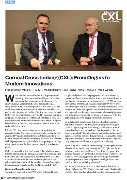
A sua descoberta veio de uma fonte inesperada. Inspirado pela utilização de luz ultravioleta (UV) para endurecer resinas dentárias, Seiler imaginou uma abordagem semelhante para a córnea. Combinando a luz UV-A com riboflavina (vitamina B₂) como fotossensibilizador, desenvolveram uma técnica para induzir ligações cruzadas covalentes no colagénio do estroma, aumentando efetivamente a rigidez da córnea. Este método localizou o efeito na córnea, minimizando os danos nos tecidos circundantes.
Normalização da terminologia e dos protocolos
O procedimento, inicialmente conhecido por vários nomes, como "reticulação do colagénio da córnea" e "C3-R", foi padronizado como "reticulação da córnea (CXL)" durante a Reunião de Peritos em CXL de 2008, em Zurique. O "protocolo de Dresden", o método original de efetuar o CXL, resistiu ao teste do tempo e continua a ser a norma de ouro. Estudos a longo prazo demonstraram a sua eficácia em travar a progressão do ceratocone durante mais de 15 anos.
Olhando para o futuro: O futuro do CXL
Embora as técnicas tradicionais de CXL continuem a ser eficazes, o futuro reserva-nos avanços promissores. Uma dessas inovações é o CXL de dois fótons, que utiliza lasers de femtosegundo para reticular seletivamente o tecido da córnea em pontos tridimensionais específicos. Esta tecnologia poderá expandir as aplicações do CXL para além das doenças ectásicas, oferecendo possivelmente novos tratamentos para a miopia e o astigmatismo.
Conclusão
Ao celebrarmos 25 anos de CXL, é evidente que o campo continua a evoluir. Inovações como o CXL de dois fótons e as cirurgias refractivas terapêuticas personalizadas estão a expandir os horizontes do que é possível no tratamento da córnea.
Melhorar a qualidade de vida através da cirurgia refractiva terapêutica
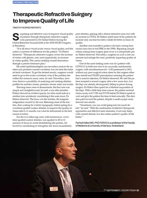
Farhad Hafezi discutiu o impacto da cirurgia refractiva terapêutica na qualidade de vida dos doentes durante a Cimeira Refractiva Global no Congresso da ESCRS de 2024 em Barcelona. Ele enfatizou a distinção entre acuidade visual e qualidade visual, defendendo tratamentos que abordam as aberrações da córnea para melhorar a função visual geral.
Estudos de casos que destacam técnicas avançadas
O Prof. Hafezi apresentou vários casos em que foram utilizadas técnicas avançadas de diagnóstico e de cirurgia:
Cicatriz após ceratite: Ao libertar a tração causada pelas cicatrizes e ao realizar uma ablação superficial guiada por uma frente de onda, foram obtidas melhorias significativas na qualidade visual ao longo de 6 a 12 meses.
Doentes com ceratocone: A combinação de uma remoção limitada de tecido através de uma ablação superficial guiada pela frente de onda com CXL imediato fortaleceu a córnea sem comprometer a integridade estrutural, melhorando a acuidade visual à distância corrigida (CDVA).
Complicações pós-PRK: Em doentes com coma horizontal elevado devido a procedimentos PRK desactualizados, a recentragem e o alargamento da zona ótica conduziram a melhorias notáveis na qualidade visual.
Uma abordagem holística à correção da visão
O Prof. Hafezi sublinhou a importância de abordar tanto a estrutura da córnea como a função visual. Ao tirar partido dos avanços no diagnóstico por imagem e no planeamento de tratamentos personalizados, os oftalmologistas podem oferecer soluções que melhoram significativamente a qualidade de vida dos doentes.