
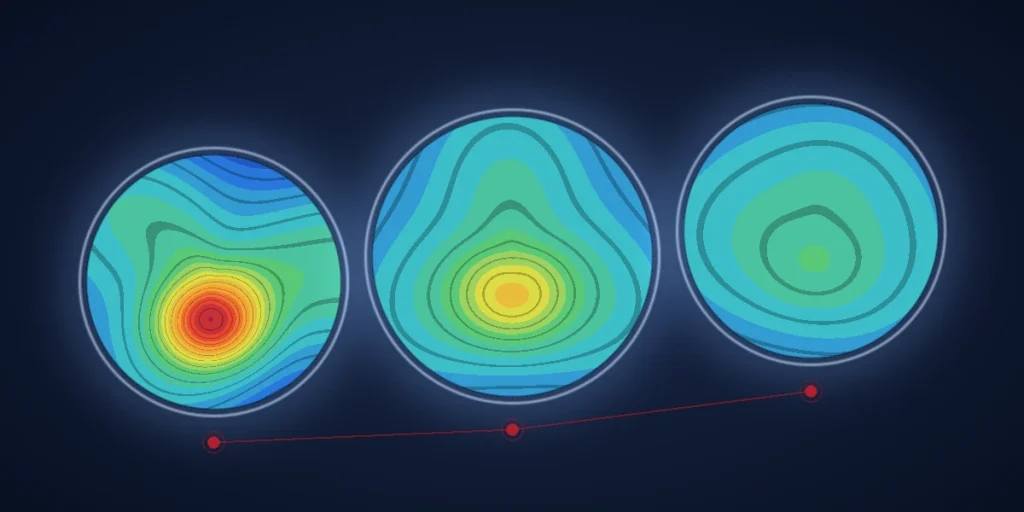
Many people with keratoconus still read the eye chart reasonably well. Yet they describe a veil over their vision, double outlines, halos and glare at night. The chart measures sharpness; it does not capture the distorted optics behind those complaints. In the September 2026 cornea issue of Der Augenspiegel, Prof. Farhad Hafezi sets out ELZA’s answer: stepwise visual rehabilitation, which moves from stabilising the cornea to reshaping it and finally fine-tuning its optics.
Why glasses are often not enough
Keratoconus is a progressive thinning and weakening of the cornea that usually begins in the second decade of life. As the front and back surfaces of the cornea become irregular, they create higher-order aberrations. These optical errors, above all vertical coma, cannot be corrected with glasses or soft contact lenses. Scleral or rigid contact lenses can often mask these errors. However, not everyone tolerates them long term, and they do nothing for the underlying biomechanical weakness.
Conservative measures come first: better optical correction, treating eye allergies and avoiding eye rubbing. When they are not enough, several surgical options are now available. Prof. Hafezi notes that one question comes up again and again in clinic: what is the current standard of care? “The honest answer is: it depends.”
Stepwise visual rehabilitation, the ELZA pyramid
ELZA organises these options as a pyramid of three steps, used alone or in combination:
- Step 1, stabilisation: halting progression with corneal cross-linking (CXL).
- Step 2, first visual improvement: regularising the shape of the cornea with corneal allogenic intrastromal ring segments (CAIRS or ECO-CAIRS) or with ELZA-PACE.
- Step 3, fine-tuning: reducing the remaining higher-order aberrations with wavefront- or topography-guided transepithelial PRK.
Which step suits which eye depends on topography, tomography, corneal thickness, biomechanical imaging and the patient’s own wishes. A guiding principle is to remove as little corneal tissue as possible. Donor-tissue ring segments can be removed or exchanged, whereas cross-linking and laser ablation change the cornea permanently. In this way, the cornea stays “available” for later steps.
Step 1, stabilising the cornea
Cross-linking stiffens the cornea using riboflavin (vitamin B2) drops and ultraviolet light. Since the Dresden protocol was first described in 2003, it has become the standard treatment for progressive keratoconus. Over two decades it has also grown from a fixed recipe into a platform that can be personalised. The article highlights three current directions. The ELZA-sub400 protocol treats corneas thinner than 400 µm. Modern epi-on protocols leave the surface layer intact. Finally, high-fluence cross-linking delivers 10 J/cm². In laboratory studies, epi-on cross-linking with a modified penetration enhancer stiffened the cornea as much as accelerated epi-off cross-linking. Whether this translates into the same long-term clinical effect still needs to be studied. ELZA has since reported 1-year clinical results for its epi-on protocol.
Step 2, reshaping the cornea
ELZA-PACE (PTK-assisted customised epi-on CXL) aims to regularise the cornea as well as stabilise it, without removing stromal tissue. It combines four gradients. First, a laser removes a thin layer of epithelium, about 8–10 µm, over the steepest part of the cone. As a result, riboflavin and oxygen reach the stroma more easily there. Finally, pulsed high-fluence UV light with a central boost concentrates cross-linking where the cornea is weakest. The clinical results of ELZA’s patient cohort are currently under peer review. In Prof. Hafezi’s words, PACE moves cross-linking from a purely stabilising procedure to “a first building block of visual rehabilitation”.
When PACE does not regularise the cornea enough, ring segments come into play. Plastic (PMMA) ring segments have been used for over 20 years. However, they come in fixed lengths and can occasionally migrate or extrude. CAIRS, introduced by Soosan Jacob in 2018, use segments cut from donor cornea instead. Because they are made of collagen, they integrate biologically and can sit in shallower layers for a stronger reshaping effect. ECO-CAIRS goes one step further: the donor segments are cross-linked at ultra-high fluence outside the eye before implantation. The stiffened segments hold their shape during implantation and fit into the cornea more easily, with less swelling. Whether this gives more predictable long-term results than untreated CAIRS still needs larger comparative studies. In ELZA’s experience so far, ECO-CAIRS can be considered particularly in moderate to advanced keratoconus. Regularising the cornea before cataract surgery can also make lens power calculations more reliable.
Step 3, fine-tuning the optics
On a stable and, ideally, already regularised cornea, wavefront-guided transepithelial PRK can then refine the remaining visual errors. The goal is not to correct the full prescription but to reduce higher-order aberrations, especially vertical coma. Prerequisites include documented stability, sufficient corneal thickness, a limited planned tissue removal and a suitable cone shape. At the ELZA Institute, this step uses a SCHWIND AMARIS excimer laser with an integrated wavefront-guided algorithm. Newer algorithms also take the back surface of the cornea into account and may help avoid unnecessary tissue removal. Their added benefit still needs to be confirmed in prospective studies.
Options beyond a corneal transplant
Prof. Hafezi sums up stepwise visual rehabilitation as a graded “both-and” rather than an “either-or”: stabilise with cross-linking, reshape with ECO-CAIRS or ELZA-PACE, and fine-tune with wavefront-guided PRK, keeping each step as tissue-sparing as possible. His message to colleagues is that even advanced keratoconus now has realistic options beyond a full-thickness corneal transplant. In addition, early referral to a specialist centre preserves those options for both the eye and the patient.
The full article on stepwise visual rehabilitation appears in Der Augenspiegel, September 2026, pages 30–33. More on ELZA’s approach is available on its page about improving vision in keratoconus and in an earlier overview of the ELZA approach to rehabilitating keratoconus corneas. Patients who want to know which step might suit them can book a consultation at the ELZA Institute.
