Cross-linking has been halting keratoconus since the early 2000s. The field is therefore old enough to face an uncomfortable question: does the effect hold? Ten year cross linking outcomes opened the free-paper session on advanced cross-linking in keratoconus on Sunday 13 September. Three of the eighteen papers in that session came from ELZA.
Ten year cross linking outcomes from the original protocol
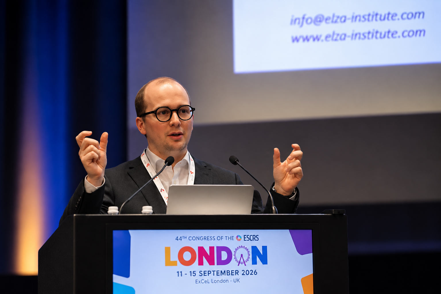
Dr Michalina Furrer presented first, at 08:30. Her subject was eyes treated with the original Dresden protocol, during the early years of cross-linking in Zurich. Corneal cross-linking (CXL) stiffens a weakened cornea using riboflavin – vitamin B2 drops – and ultraviolet light. The Dresden protocol is the version everything else is measured against. The epithelium is removed, and the cornea then receives 3 mW/cm² for 30 minutes, a total of 5.4 J/cm².
Of 418 treated eyes, 51 had complete ten-year data. After excluding eyes that had undergone further procedures, 38 were analysed. Mean follow-up was 10.9 years. Maximum keratometry, or Kmax, is a measure of how steep the cornea has become. It fell by 1.30 D. Corrected distance visual acuity improved by 0.15 logMAR. Just over half the eyes flattened by more than 1 D, 39% stayed within 1 D of baseline, and 8% progressed. Overall long-term stabilisation was 90%. One eye needed repeat cross-linking, and two went on to keratoplasty. No late infections, limbal stem cell problems or visually significant scarring were seen.
The honest caveat sits in the numbers themselves. Only 51 of 418 eyes returned with complete ten-year data. That is a familiar problem in long-term corneal follow-up, and it means the analysed group may not represent the whole cohort. Even so, these ten year cross linking outcomes are among the longest published for the Dresden protocol.
When cross-linking goes wrong
At 08:50, Léonard Kollros presented a case series the field needs more of. Fifteen eyes of eleven patients developed central toxic keratopathy after standalone epithelium-off cross-linking. All were treated at an external centre, and later sought assessment at ELZA independently. Central toxic keratopathy is a non-inflammatory clouding and thinning of the central cornea.
Ten eyes had dense central stromal scarring, while five had central haze. Corrected acuity ranged from 20/20 to 20/200. Imaging showed marked central thinning, below 300 µm in severe cases. Hyperreflectivity ran through the full stromal thickness. Several cases involved accelerated protocols, or substantial thinning during surgery after dextran-containing riboflavin.
The authors point to three contributing factors: excessive ultraviolet exposure, intraoperative thinning, and unregulated riboflavin formulations. They argue for strict ultraviolet calibration, intraoperative pachymetry, and careful choice of riboflavin. Because the cases were treated elsewhere and referred in, the series cannot give an incidence rate. It documents what the complication looks like, which is a different and still useful thing.
Ring segments that hold their shape
At 09:20 Kollros returned with laboratory work on corneal allogenic intrastromal ring segments, or CAIRS. These are arcs of donor corneal tissue, implanted into the stroma to flatten a cone. Forty-eight porcine corneas were used to prepare segments. The segments were randomised to no ultraviolet treatment, to 30 J/cm², or to 60 J/cm² of extracorporeal cross-linking. All then went through a standardised dehydration and rehydration cycle.
Both cross-linked groups dehydrated significantly more than controls, and in addition rehydrated significantly less. There was no difference between 30 J/cm² and 60 J/cm². In practical terms, the segments end up thinner and slower to re-expand. That should make them easier to handle, and extend the window a surgeon has to implant them. This is ex vivo work in pig tissue, and the authors say plainly that in vivo studies are needed.
Chairing, and a customised protocol on the poster floor
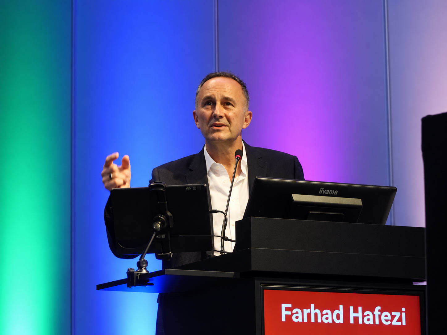
Prof. Farhad Hafezi chaired two presented poster sessions during the day. The first covered laser vision correction at 09:15, the second corneal cross-linking at 16:00. Both sessions ran in the exhibition area. In the second, at 16:35, he presented one and two-year outcomes of ELZA-PACE in 135 keratoconus eyes.
ELZA-PACE removes epithelium selectively over the tip of the cone. That creates a small epi-off zone inside an otherwise epi-on cornea. Ultraviolet energy is then delivered unevenly, with the highest fluence over the ectatic region. Reported flattening reached up to 12 D of maximum keratometry in selected cases, without endothelial decompensation or clinically relevant haze. Cone shape, age and disease activity all influenced how much flattening occurred. In other words, results vary by eye, and the selected cases are not the average.
The ELZA Institute performs corneal cross-linking for keratoconus at its clinics in Dietikon and Zurich, including ELZA-PACE. Session details are published by the European Society of Cataract and Refractive Surgeons.