A cornea can look normal and still not behave normally. Closing that gap between shape and behaviour is the work of corneal biomechanics keratoconus research. It ran through everything ELZA presented on Saturday 12 September at the 44th ESCRS Congress in London.
Why corneal biomechanics matters in keratoconus
Keratoconus is a weakening disease. The cornea, the clear dome at the front of the eye, loses stiffness. As a result, it slowly bulges into a cone, and vision blurs. For decades, the only way to detect the disease was to measure corneal shape. Shape, after all, is what cameras can see. The problem is that shape changes late. By the time a cornea looks abnormal, tissue has already been lost.
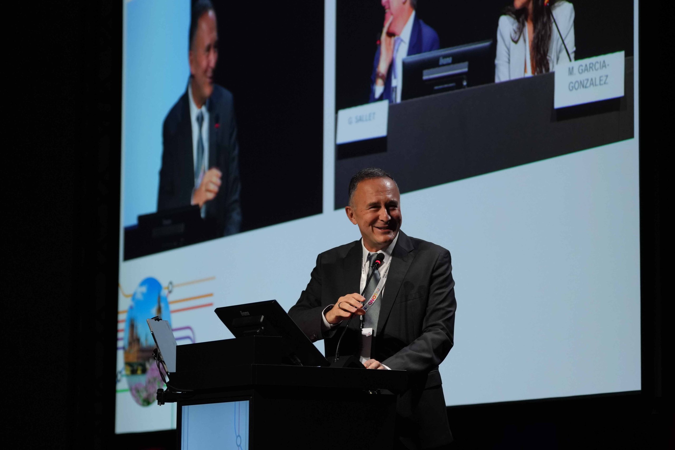
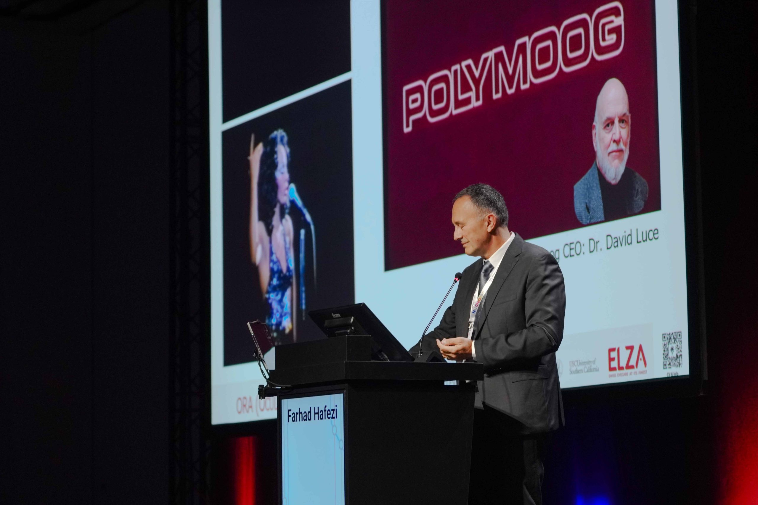
Biomechanical measurement takes a different route. A high-speed camera films the cornea as a puff of air deforms it. How the tissue moves reveals how stiff it is. Prof. Farhad Hafezi set out that case at 09:25, in the Refractive Surgery Didactic Course in Victoria Room 1/2.
What corneal biomechanics keratoconus screening found in children
The argument became concrete at 14:20, in the presented poster session on keratoconus and secondary ectasia. There Prof. Hafezi reported on the children of keratoconus patients. The study looked at 322 eyes of 161 children, each with at least one affected parent. All were imaged with Scheimpflug tomography. In addition, 95 had biomechanical assessment.
Across the whole group, 31.7% of the children showed tomographic abnormalities. A further 11.2% reached values usually considered pathological. In the subgroup that received both tests, 65.3% had at least one abnormal biomechanical index. Expert clinical assessment identified keratoconus outright in 18.6% of the children, and keratoconus suspect in another 13.7%. Both kinds of abnormality increased markedly with age.
Those rates sit well above what is expected in the general paediatric population. The practical implication is twofold. First, the children of keratoconus patients are a screening population. Second, paediatric normative data are needed to read their results properly, because adult thresholds were never designed for developing eyes.
Reshaping the cornea without removing tissue
The afternoon symposium in George V Room 1 asked a pointed question. Can penetrating keratoplasty for keratoconus be made obsolete? In other words, can full-thickness corneal transplantation be avoided in most cases? Three ELZA talks addressed different parts of that question.
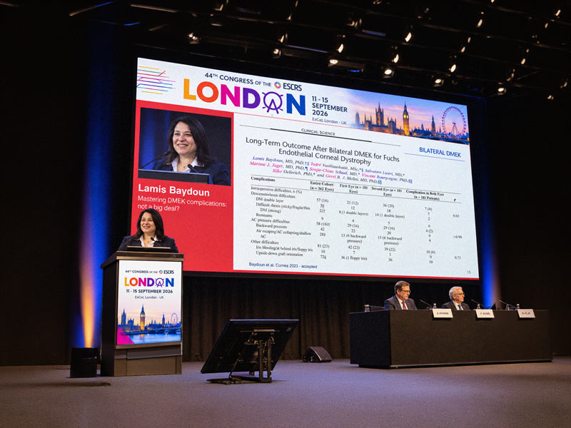
At 14:30 Dr Lamis Baydoun spoke on Bowman layer transplantation. The technique inserts a thin donor membrane into the stroma to flatten an advanced cone. At 14:55 Prof. Hafezi presented epithelial map-guided customised epi-on cross-linking, the protocol ELZA calls ELZA-PACE. At 15:40 Dr Emilio A. Torres-Netto presented ECO-CAIRS, in which donor tissue ring segments are cross-linked outside the eye before implantation.
What links the three is a shared premise. A weakened cornea can be reinforced and regularised rather than replaced. All three approaches are additive or in situ, and none removes stromal tissue. Each therefore keeps the corneal biomechanics keratoconus question open in a useful way. The tissue that remains is the tissue being strengthened.
Bullous keratopathy, a common problem with an unglamorous name
Earlier, at 08:00, Dr Baydoun co-chaired a clinical research symposium on bullous keratopathy. The condition is corneal swelling caused by endothelial pump-cell failure, most often after cataract surgery. It is one of the commonest reasons for a corneal transplant worldwide. Even so, it receives a fraction of the attention that keratoconus does.
The ELZA Institute treats keratoconus at its clinics in Dietikon and Zurich. It offers ELZA-PACE customised cross-linking and ECO-CAIRS there. Session details are published by the European Society of Cataract and Refractive Surgeons.