Congresses front-load their subspecialty days. For anyone who works on the cornea, the ESCRS 2026 Cornea Day is where the week begins in earnest. It ran on Friday 11 September in Victoria Room 1/2 at ExCeL London. Prof. Farhad Hafezi, Chief Medical Officer at the ELZA Institute, co-chaired its keratoconus and crosslinking session with Prof. Jesper Hjortdal.
Cross-linking that aims to do more than stop the disease
Keratoconus weakens the cornea, the clear dome at the front of the eye. Over time the cornea bulges into a cone, and vision blurs. Corneal cross-linking (CXL) halts that process. It combines riboflavin – vitamin B2 drops – with ultraviolet light, which stiffens the collagen inside the tissue. CXL has been standard care for two decades. For most of that time, however, the goal was simply to stop the disease getting worse.
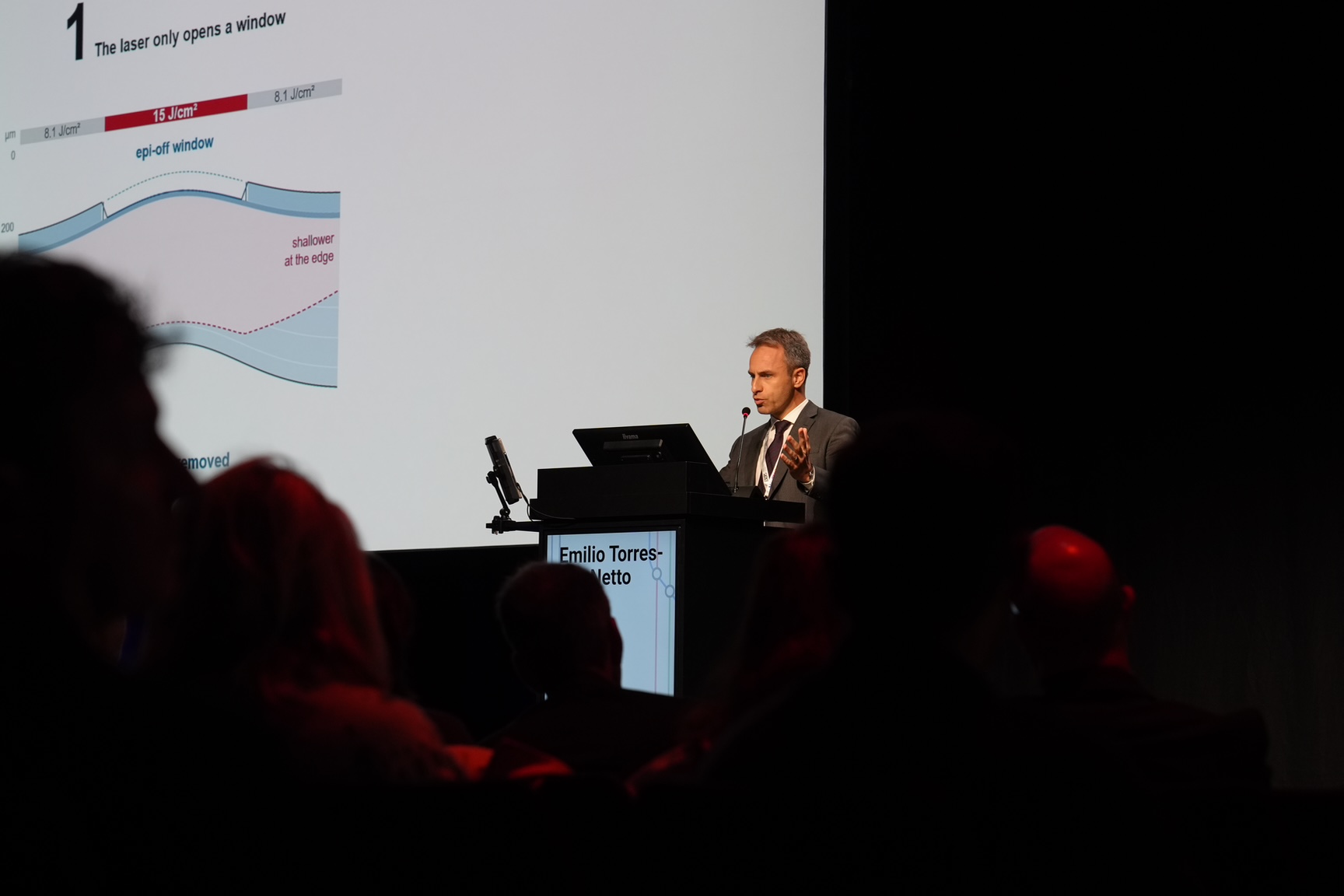
Dr Emilio A. Torres-Netto argued for a larger ambition at 15:10, in a talk titled CXL to Improve Vision. The premise is straightforward. A cornea that can be stiffened can also be reshaped, given enough control over where the energy goes. Customised protocols therefore concentrate ultraviolet fluence over the cone itself, instead of treating the cornea uniformly. As a result, the steepest part flattens more than the rest. Because the surrounding tissue is mechanically coupled to it, the whole surface becomes more regular. No tissue is removed.
What the ESCRS 2026 Cornea Day session covered
The ESCRS 2026 Cornea Day session ranged across the keratoconus treatment ladder. It moved from cross-linking through intrastromal ring segments to transplantation. That progression matters clinically. Each rung is chosen on how much cornea is left to work with. It also depends on how much vision has already been lost.
Corneal transplantation on the Friday programme
Earlier in the afternoon, Dr Lamis Baydoun spoke in the Cornea Surgery session at 12:45. Her subject was the endothelium and anterior chamber intraocular lenses. The endothelium is the single layer of pump cells on the back of the cornea. It keeps the tissue clear. When it fails, the cornea swells and vision clouds.
DMEK, or Descemet membrane endothelial keratoplasty, replaces only that layer. Older techniques replace the full thickness of the cornea instead. Recovery is therefore faster, and rejection rates lower. In addition, Dr Baydoun taught the intermediate DMEK wet lab at 15:00, where surgeons practise graft handling on donor tissue. Wet labs rarely make congress write-ups. They are, however, where techniques actually spread between surgeons.
Glaucoma Day and the case for debate
Prof. Kaweh Mansouri opened ELZA’s Friday at 09:30, with a contribution to Mind the Gap, a session on the deciding moments in glaucoma care. He then chaired the debate session Modern Standards in Glaucoma Care at 10:00, in George V Room 2. Glaucoma damages the optic nerve, usually alongside raised pressure inside the eye. That damage is largely irreversible. Consequently the arguments are about when to intervene, rather than whether.
Five ELZA e-posters, available from the opening day
The congress e-posters went live on demand from Friday, and five of them come from ELZA. Two deal with cross-linking directly: repeat treatment after a first CXL failed, and imaging agreement during follow-up. A third reports biomechanical screening in Uzbek schoolchildren. A fourth covers keratoconus prevalence in Russia, from the K-MAP study. The fifth reports twelve-month outcomes of a supraciliary glaucoma drainage device. Delegates can read all five in their own time during the week.
The ELZA Institute treats keratoconus at its clinics in Dietikon and Zurich. It also performs corneal cross-linking, corneal surgery and glaucoma care there. The full programme is published by the European Society of Cataract and Refractive Surgeons.